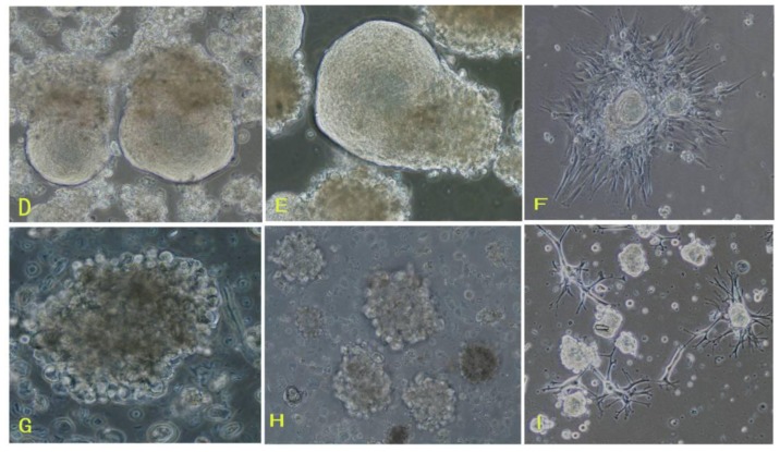
Figure 1.
Morphologies of cells from three subtypes of meningiomas. (A–C) Cells from fiber meningiomas (A: primary spheres 200×, B: the third spheres 200×, C: adherent sphere 100×); (D–F) Cells from hemangiopericytoma meningiomas (D: primary spheres 200×, E: the third spheres 200×, F: adherent sphere 100×); (G–I) Cells from epithelial meningiomas (G: primary spheres 200×, H: the third spheres 200×, I: adherent sphere 100×).


