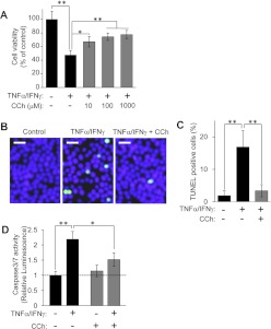
Fig. 1.
CCh protects HSG cells from proinflammatory cytokine-induced apoptosis. A, HSG cells were treated with or without various concentrations of CCh (10–1000 μM), in the absence or presence of TNFα (50 ng/ml)/IFNγ (10 ng/ml), and were incubated for 56 h. Cell viability was determined as described under Materials and Methods and is shown as a percentage of the viability of cells grown in medium B. Values represent the mean ± S.D. of four cultures. *, p < 0.05; **, p < 0.01, values differ significantly (t test). B to D, HSG cells were treated with or without 100 μM CCh, in the absence or presence of TNFα (50 ng/ml)/IFNγ (10 ng/ml), and were incubated for 40 h (B and C) or 24 h (D). B and C, TUNEL-positive apoptotic cells (green) are shown under each set of conditions (B), and the graph shows the percentage of TUNEL-positive apoptotic cells (C). Values represent the mean ± S.D. of three cultures. **, p < 0.01, values differ significantly (t test). Similar results were obtained from three experiments. D, caspase 3/7 activity was indicated by luminescence activity, as described under Materials and Methods. Values represent the mean ± S.D. of three cultures. *, p < 0.05; **, p < 0.01, values differ significantly (t test).

