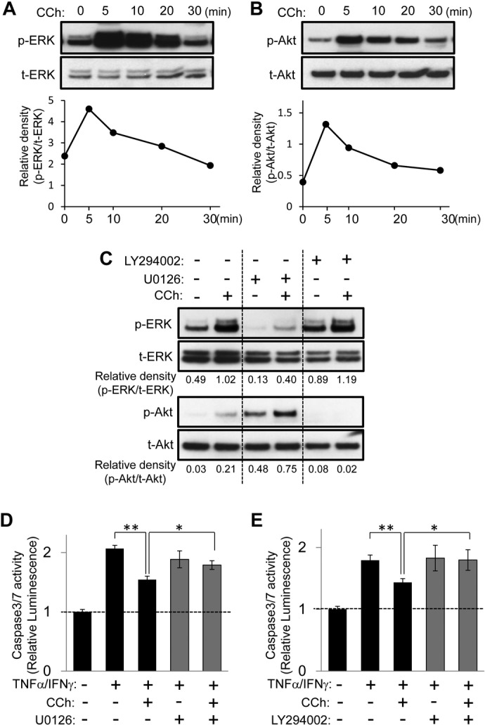
Fig. 2.

ERK and Akt signaling pathways are essential for CCh-promoted cytoprotection against apoptotic challenges in HSG cells. A and B, HSG cells were exposed to CCh (100 μM) for the indicated times. The phosphorylated (p) and total (t) ERK (A) and Akt (B) levels were analyzed through immunoblotting. C, HSG cells were pretreated with or without U0126 (2.5 μM) or LY294002 (2.5 μM) for 30 min and then were exposed to CCh (100 μM) for 10 min. The phosphorylated and total ERK and Akt levels were analyzed through immunoblotting. Quantification of the band density was performed through densitometric scanning of each band by using National Institutes of Health ImageJ software. D and E, HSG cells were pretreated with or without U0126 (2.5 μM) (D) or LY294002 (2.5 μM) (E) for 30 min. The cells then were treated with or without 100 μM CCh, in the absence or presence of TNFα (50 ng/ml)/IFNγ (10 ng/ml), and were incubated for 24 h. Caspase 3/7 activity was indicated by luminescence activity, as described under Materials and Methods. Values represent the mean ± S.D. of three cultures. *, p < 0.05; **, p < 0.01, values differ significantly (t test).
