Figure 2.
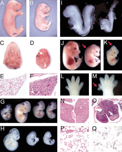
Inactivation of Ovca1 or Ovca1-2 causes developmental abnormalities. Gross morphology of E18.5 (A) wild-type and (B) Ovca1-2-/- pups. Ventral view of palate of E18.5 (C) wild-type and (D) Ovca1-2-/- mice, showing cleft palate in the mutant (arrow). (E,F) H&E-stained histological sections of lung from E18.5 wild-type (E) and Ovca1-2-/- (F) pups. (G) E10.5-E11.5 embryos. E10.5 wild-type embryo (left); E11.5 wild-type embryo (second from left); two E11.5 Ovca1-2-/- embryos (right). (H) E9.5 embryos. Wild-type embryo (left); two Ovca1-2-/- embryos (right). (I) E8.5 embryos. Wild-type embryo (left); Ovca1-2-/- embryo (right). (J) E14.5 embryos. Wild-type embryo (left); Ovca1-/- embryo (right) with edema (arrow). (K) E13.5 Ovca1-2-/- embryo with midbrain exencephaly (arrow). (L) Hindlimb of an E14.5 wild-type embryo. (M) Preaxial polydactyly (arrow) in right hindlimb of E14.5 Ovca1-2-/- embryo. (N,O) H&E-stained histological sections of E16.5 fetal liver from wild-type (N) and Ovca1-2-/- (O) embryos. The mutant liver is highly basophilic and has a focal area of necrosis (arrow). (P,Q) Representative Wright-Giemsa-stained blood smears of E16.5 wild-type (P) and Ovca1-2-/- or Ovca1-/- (Q) embryos. Note that many of the mutant erythrocytes are nucleated.

