Figure 6.
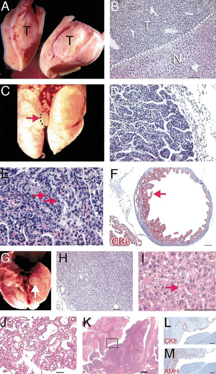
Spontaneous tumor formation in Ovca1-2+/- and Ovca1+/- mice. (A) Tumor mass (T) found in the liver of an Ovca1-2+/- mouse. (B) H&E-stained section of tumor shown in A. Hepatocellular adenoma (T) showing basophilic, focal eosinophilic, and clear cell appearance with compression of the normal hepatic parenchyma (N). Dashed line indicates the boundary between tumor and normal hepatic parenchyma. Scale bar, 100 μm. (C) Tumor mass (arrow) found in the lung of an Ovca1-2+/- mouse. (D) H&E-stained section of bronchiolo-alveolar carcinoma (T) with mixed papillary and solid patterns shown in C. Scale bar, 100 μm. (E) Higher magnification of D, showing mitotic figures (arrowheads). (F) Cystadenoma found in the ovary of an Ovca1-2+/- mouse at 80 wk. Papillary-like epithelium immunostained for cytokeratin 8 (CK8, red) projects into the cyst, which is lined with tall columnar cells (arrow). Scale bar, 400 μm. (G) Tumor mass (arrow) found in a ventral lung lobe of an Ovca1+/- mouse at 78 wk of age. (H) H&E-stained section of tumor shown in G, showing a predominantly glandular arrangement of the tumor cells. (I) H&E-stained section of hepatocellular ademoma of an Ovca1+/- mouse at 66 wk of age. There is an abnormal presence of many hyaline bodies (arrow). Scale bar, 100 μm. (J) Mammary gland tumor found in an Ovca1+/- female at 70 wk of age. Adenocarcinoma type B was indicated because of collagenousstroma and glandular components consisting of focal cystic, hemorrhagic, and papillary growth patterns. Scale bar, 100 μm. (K) Tubular adenoma found in the ovary of an Ovca1+/- female at 82 wk of age. (Box) Region shown in L and M. Scale bar, 400 μm. (L) The neoplastic cells were lined by surface mesothelial cells which expressed CK8 (red). Scale bar, 100 μm. (M) AMH (red) was detected focally in the tumor. Scale bar, 100 μm.

