Figure 7.
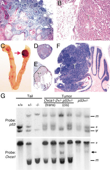
Interaction between Ovca1 and p53 mutations for tumor formation. (A) H&E-stained section of a representative mammary gland adenosquamous carcinoma was found in two Ovca1-2+/- p53+/- mice at 38 and 78 wk of age. There are keratinized well-differentiated squamous cells surrounded by the glandular elements of the mammary gland. Scale bar, 100 μm. (B) H&E-stained section shows a bronchiolo-alveolar carcinoma with a papillary growing pattern in an Ovca1-2+/- p53+/- male mouse at 80 wk of age. Scale bar, 100 μm. (C) Reproductive tract from an Ovca1-2+/- p53-/- female mouse at 33 wk of age. One ovary (arrow) shown at the right is enlarged and hemorrhagic. H&E-stained sections of ovaries shown in C with normal histology (D) and a hemangiosarcoma immunostained with CD34 (brown; E). (F) H&E-stained section of a brain tumor (T) showing a transition from the cerebellar granular layer to a neoplastic appearance reminiscent of human medulloblastoma, replacing the cerebellar folia. (G) Southern blot analysis of p53 and Ovca1 in tumors from Ovca1-2+/- p53+/- and p53+/- mice. Tail DNA from wild-type (+/+), cis Ovca1-2+/- p53+/- (+/-), and Ovca1-2-/- p53-/- (-/-) mice. Genomic DNA from individual tumors, including trans Ovca1-2+/- p53+/-, cis Ovca1-2+/- p53+/-, and p53+/-. (m) Mutant allele; (+) wild-type allele; (ψ) p53 pseudogene. One cis Ovca1-2+/- p53+/- tumor shows LOH of the p53 locus and an additional minor band (arrow), suggesting a rearrangement of the Ovca1 locus in a subset of cells in the tumor.

