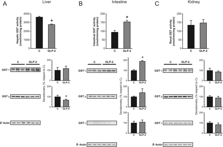
Fig. 2.
Effect of GLP-2 on activity and expression of hepatic, intestinal, and renal GST. GST activity toward CDNB (top) and Western blot studies of main GST classes (bottom) were performed in cytosol isolated from liver (A), jejunal mucosa (B), and renal cortex (C). For Western blots, equal amounts of protein (10 μg) were loaded in all lanes. Expression of GST-Alpha, -Mu, and -Pi was calculated relative to β-actin expression. Uniformity of protein loading and transfer from gel to PVDF membrane was controlled with Ponceau S. Data on densitometric analysis are presented as percentages relative to control, considered as 100%, and were expressed as means ± S.D. of six rats per group. Hepatic GST-Pi was not detected in either control or GLP-2 groups.*, significantly different from control (C), P < 0.05.

