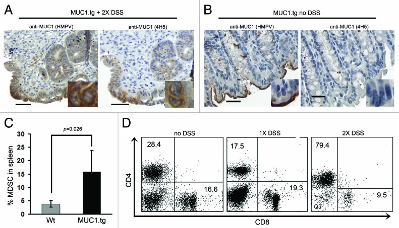
Figure 2.DSS-induced colitis in MUC1tg mice is characterized by increased expression of abnormal MUC1, increased percentage of MDSC and loss of CD8+ T cells. Immunostaining of colon tissue sections from MUC1.tg mice that received two cycles of DSS (A) and no DSS control mice (B). HMPV antibody stains all MUC1; 4H5 antibody stains hypoglycosylated MUC1. Scale bars are 100 µm. Inserts were taken with 100x oil-immersion objective. (C) Isolated spleen cells from Wt (n-4) and MUC1.tg (n = 4) mice were stained for Gr-1 and CD11b expression and analyzed by flow cytometry. p was determined by two-tailed unpaired t-test. (D) Isolated spleen cells from control MUC1.tg mice that received no DSS, MUC1.tg mice that received one cycle of DSS, and MUC1.tg mice that received two cycles of DSS were labeled for CD4 and CD8 and analyzed by flow cytometry. Representative dot plots from two separate experiments.

An official website of the United States government
Here's how you know
Official websites use .gov
A
.gov website belongs to an official
government organization in the United States.
Secure .gov websites use HTTPS
A lock (
) or https:// means you've safely
connected to the .gov website. Share sensitive
information only on official, secure websites.
