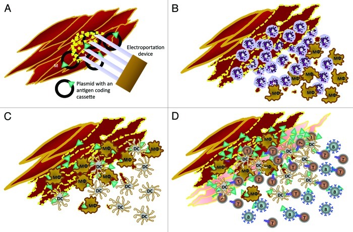
Figure 1. Cellular events following intramuscular electroporation of 50 µg of plasmid in 20 µl of saline through Cliniporator (Igea, Carpi, Italy). Insertion of the electroporator needles into the quadriceps muscle of the mouse and delivery of two low voltage pulses of 150V of 25 ms with a 300 µs interval (A). One-6 h later the damaged myofibers and cell debris (dotted yellow) are surrounded by polymorphonuclear (N) and mononuclear leukocytes (MΦ) (B). One or two days later mature and differentiated tissue macrophages (MΦ) and dendritic cells (DC) progressively become prominent among inflammatory cells infiltrating the numerous necrotic myofibers (C). On the third-fourth day from the electroporation, intact and regenerating muscular fibers (pale red) are overexpressing the protein encoded by the plasmid (green triangles), while the area is being infiltrated by B (B), T (T) and dendritic cells (DC). Interestingly, CD11b+ macrophages and CD11c+ DC and, later, CD4+ T cells are often in direct contact with antigen expressing muscle cells or antigen expressing fragments and each other (D).

An official website of the United States government
Here's how you know
Official websites use .gov
A
.gov website belongs to an official
government organization in the United States.
Secure .gov websites use HTTPS
A lock (
) or https:// means you've safely
connected to the .gov website. Share sensitive
information only on official, secure websites.
