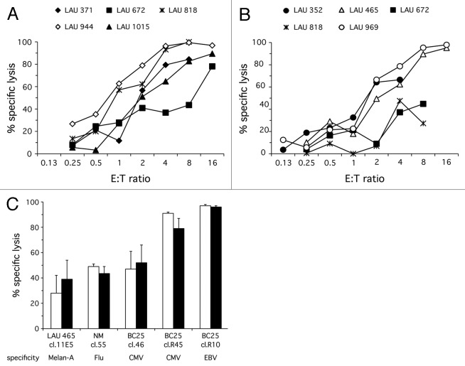
Figure 1. Melan-A-specific T-cells from both PBMC and TILN of melanoma patients exert strong cytotoxicity directly ex vivo. Melan-A-specific CD8+ T-cells were FACS purified from PBMC [(A); n = 5] and TILN [(B); n = 5] of melanoma patients using pMHC multimers, and incubated for 4 h with an equal number of Melan-A-pulsed T2CMTMR-lo and HIV-pulsed T2CMTMR-hi at the indicated E:T ratios. All conditions were measured in triplicates. A semi-paired permutation test yielded no statistically significant difference between the cytolytic capacity of cells from PBMC and TILN (p = 0.2). (C) Pre-incubation with pMHC multimers does not significantly affect the lytic activity of CD8+ CTL clones. CTL clones specific for either Melan-A, Flu, CMV or EBV were left untreated (white bars) or incubated with relevant pMHC multimers (black bars) prior to incubating them for 4 h with an equal number of relevant-peptide-pulsed T2CMTMR-lo and irrelevant-peptide-pulsed T2CMTMR-hi. E:T = 4 was used for all clones except NM cl.55, which was assayed at E:T = 1. Mean cytolytic activity and SD of quadruplicates are shown for each clone. A paired, two-tailed t-test showed no difference between pMHC multimer treated or untreated cells (p = 0.94).

An official website of the United States government
Here's how you know
Official websites use .gov
A
.gov website belongs to an official
government organization in the United States.
Secure .gov websites use HTTPS
A lock (
) or https:// means you've safely
connected to the .gov website. Share sensitive
information only on official, secure websites.
