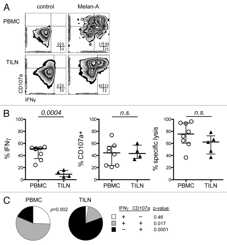
Figure 3. TILN-derived melanoma-specific CD8+ T-cells exhibit decreased IFNγ production, but comparable degranulation to their PBMC-derived counterparts. (A) Representative dot plots for degranulation and IFNγ production by PBMC- (LAU 944) and TILN-derived (LAU 465) Melan-A-specific CD8+ T-cells. (B) Frequencies of Melan-A-specific T-cells positive for IFNγ, CD107a at E:T = 0.5. and % specific lysis at E:T = 4 is indicated. Data are means of triplicates. E:T Data for PBMC and TILN were compared using unpaired, two-tailed t-tests. (C) Pies show the relative proportion of Melan-A-specific CD8+ T-cells positive for either IFNγ and/or cell surface CD107a expressing either combination of these markers.

An official website of the United States government
Here's how you know
Official websites use .gov
A
.gov website belongs to an official
government organization in the United States.
Secure .gov websites use HTTPS
A lock (
) or https:// means you've safely
connected to the .gov website. Share sensitive
information only on official, secure websites.
