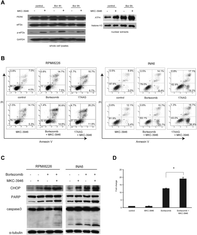
Figure 4.
MKC-3946 enhances ER stress-mediated apoptosis induced by bortezomib or 17-AAG. (A) RPMI 8226 cells were treated with bortezomib (Bor; 10nM) in the presence or absence of MKC-3946 (10μM) for the indicated times. Whole-cell lysates and nuclear extracts were subjected to Western blotting using PERK, eIF2α, phospho-eIF2α (p-eIF2α), and ATF4 Abs. GAPDH and histone H3 serve as loading controls. (B) RPMI 8226 cells were treated with bortezomib (2.5nM) or 17-AAG (500nM) and INA6 cells were treated with bortezomib (2.5nM) or 17-AAG (125nM), in each case in combination with MKC-3946 (10μM) for 24 hours. Apoptotic cells were analyzed by flow cytometry using annexin V/PI staining. (C) RPMI 8226 and INA6 cells were treated with bortezomib (2.5nM) in the presence or absence of MKC-3946 (10μM) for 24 hours. Whole-cell lysates were subjected to Western blotting using anti-CHOP, PARP, caspase-3, and α-tubulin Abs. (D) RPMI 8226 cells were treated with MKC-3946 (10μM), bortezomib (10nM), or the combination for 8 hours. CHOP mRNA was determined by real-time quantitative PCR. Data represent mean ± SD fold changes relative to β-actin mRNA in triplicate samples. *P < .001.

