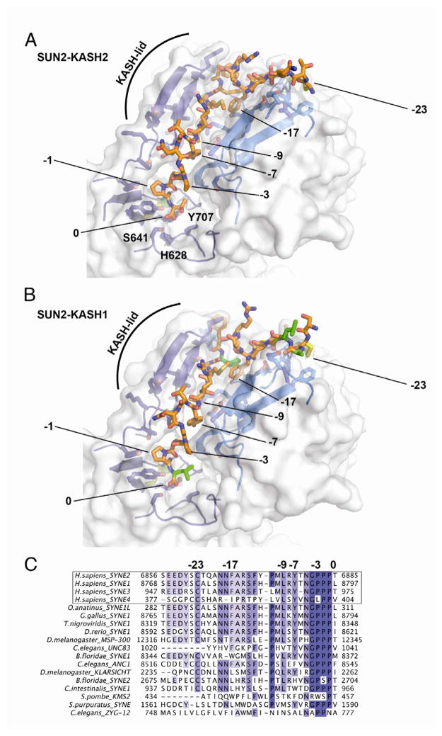
Figure 3. Details of the SUN-KASH Interaction.
(A) Close-up view of the SUN2-KASH2 interaction. Two neighboring SUN protomers are shown in two shades of blue, with the KASH2 peptides in between in orange. Surface of the SUN2 binding area is half-transparent. KASH residues crucial for interaction are numbered. ‘0’ denotes the C-terminal residue of the peptide. Pocket residues that abolish KASH-binding if mutated are labeled.
(B) Same view of the SUN2-KASH1 interaction. Residues that differ between KASH1 and KASH2 are colored in green.
(C) Multiple sequence alignment of the four identified human KASH proteins, followed by a list of KASH peptides from highly diverged eukaryotes. The numbers match residues important for SUN binding.
See also Figure S3.

