
Fig. 4.
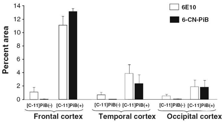
Aβ immunohistochemistry and 6-CN-PiB histofluorescence: quantitative analysis. Analysis of Aβ (6E10) immunoreactive and 6-CN-PiB amyloid deposit loads (percent area) are illustrated in frontal, temporal and occipital cortices from the [C-11]PiB(−) and [C-11]PiB(+) cases. For both markers, plaque loads in all three cortical regions of [C-11]PiB(−) case are lower than plaque load values in the [C-11]PiB(+) case. In the [C-11]PiB(+) case, 6-CN-PiB load is comparable to Aβ plaque load while in the [C-11]PiB(−) case it is substantially lower in all cortical areas