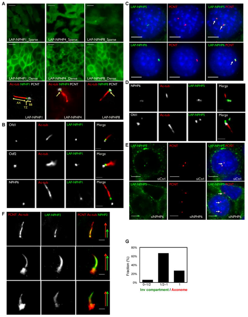
Figure 3. Localization of NPHP “1-4-8”, NPHP “5-6” and NPHP2 to the ciliary transition zone, centrosome and the inversin compartment (see also Figure S4).

A. IMCD3 cells stably expressing LAP-NPHP1 (green), LAP-NPHP4 (green) or LAP-NPHP8 (green) were immunostained for pericentrin (PCNT, white) and acetylated α-tubulin (ac-tub, red). AX: Axoneme; TZ: Transition Zone; BB: Basal Body. B. IMCD3 cells stably expressing LAP-NPHP1 (green) were immunostained for acetylated α-tubulin (ac-tub, red) and Ofd1 (white), Odf2 (white), or NPHP6 (white) C-D. NPHP5 and NPHP6 co-localize to the centrosome. C. IMCD3 cells stably expressing LAP-NPHP5 (green) or LAP-NPHP6 (green) were immunostained for pericentrin (PCNT, red). D. IMCD3 cells stably expressing LAP-NPHP5 (green) were immunostained for acetylated α-tubulin (ac-tub, red) and NPHP6 (white) or Ofd1 (white). E. Centrosomal localization of LAP-NPHP5 is disrupted upon depletion of NPHP6. IMCD3 LAP-NPHP5 (green) cells were transfected with siRNA against NPHP6 or control, and then immunostained for pericentrin (PCNT, red). Nuclei were stained with Hoechst 33258 (blue). F. NPHP5 interacting protein NPHP2 localizes to the centrosome and to the cilium. IMCD3 LAP-NPHP2 cells (green) were immunostained for pericentrin (PCNT, red) and acetylated α-tubulin (ac-tub, red). Arrows exemplify variable NPHP2/Inversin compartment extensions along the axoneme. G. Percentage of cilia with a range of “Inversin compartment / Axoneme” ratios. Scale bars, 10um (A), 5um (C) and (E), 2um (B), (D) and (F).
