Fig. 1.
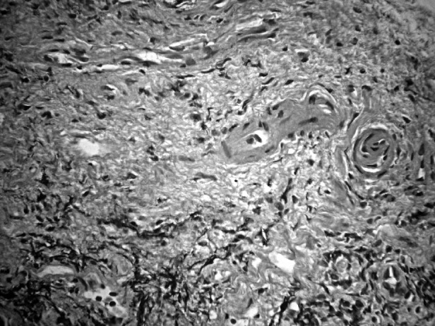
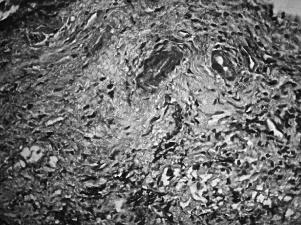
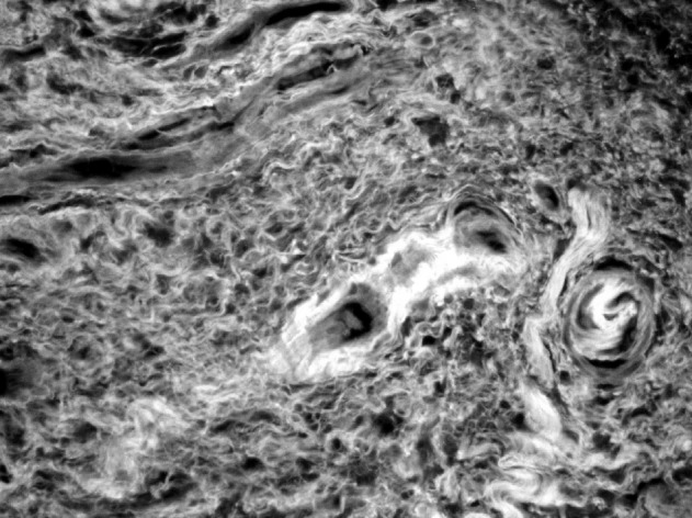
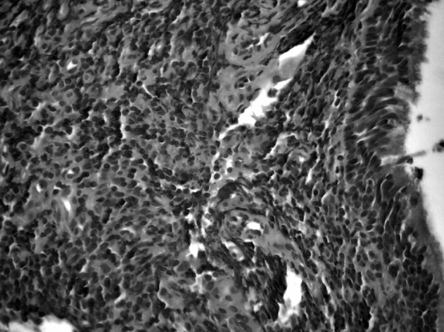
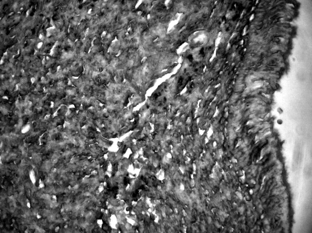
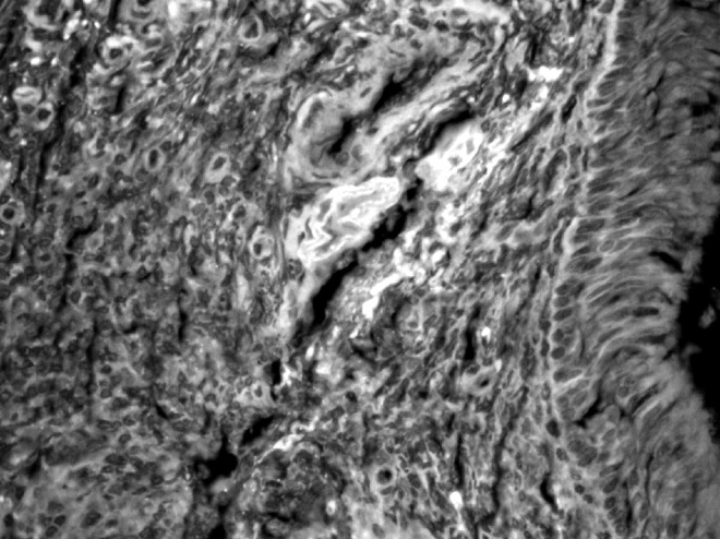
Comparison of histological features of coblation-treated (A-C) and cold curette (D-F) rhinopharynx mucosa sections. The sections from a coblationtreated patient show denuded epithelium, abundant fibrosis of the lamina propria and hyperplastic small vessels, along with focal lymphocytic infiltrates. The sections from a cold-curette treated patient show massive lymphocytic infiltration of the lamina propria with preservation of epithelial lining. A, D: H&E; B, E: Masson's trichrome stain; C, F: fluorescence microscopy observation of H&E slides (original magnification × 400).
