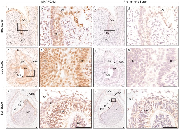
Figure 2.
Analysis of SMARCAL1 protein expression during tooth morphogenesis. (a, b) Photomicrographs of SMARCAL1 immunohistochemical staining of the bud stage of tooth development. SMARCAL1 is expressed in the cells of the oral epithelium, dental lamina, and the mesenchymal cells, which give rise to the dental papilla. (c, d) Photomicrographs of pre-immune staining of the bud stage of tooth development. The cells of the oral epithelium, dental lamina, and mesenchymal cells showed minimal non-specific staining. (e, f) Photomicrographs of SMARCAL1 immunohistochemical staining of the cap stage of tooth development. SMARCAL1 is expressed in the cells of the dental lamina, outer dental epithelium, stellate reticulum, inner dental epithelium, primary enamel knot, and dental papilla. (g, h) Photomicrographs of pre-immune staining of the cap stage of tooth development. The cells of the dental lamina, outer dental epithelium, stellate reticulum, inner dental epithelium, primary enamel knot, and dental papilla did not show non-specific staining. (i, j) Photomicrographs of SMARCAL1 immunohistochemical staining of the bell stage of tooth development. SMARCAL1 is expressed in the cells of the outer dental epithelium, stellate reticulum, stratum intermedium, inner dental epithelium, and dental papilla. (k, l) Photomicrographs of pre-immune staining of the bell stage of tooth development treated with pre-immune rabbit serum. The cells of the dental lamina, outer dental epithelium, stellate reticulum, stratum intermedium, inner dental epithelium, and dental papilla showed minimal non-specific staining. The boxed regions correspond to the higher-magnification images. Abbreviations: DL, dental lamina; DP, dental papilla; EK, primary enamel knot; IDE, inner dental epithelium; MC, mesenchymal cells; ODE, outer dental epithelium; OE, oral epithelium; SI, stratum intermedium; SR, stellate reticulum. Scale bars: 50 µm.

