Figure 8.
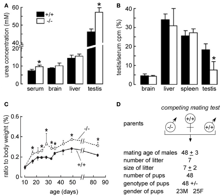
Early maturation in the male reproductive system in UT-B null mice. (A) Urea concentration in plasma and homogenized tissues. Tissues from 84-day-old mice were homogenized in water. Urea concentrations in supernatants of centrifuged homogenates were measured. (B) Kinetics of [14C] urea uptake and organ morphology in wild-type vs. UT-B null mice. After renal blood flow was blocked, a bolus of [14C] urea was injected intravenously, and the blood, brain, liver, spleen, and testis were sampled at 5 min. [14C] urea accumulation in different tissues was normalized to serum. Values are expressed as means ± SE (n = 4 mice). *p < 0.01 vs. WT mice. (C) Age-related changes in testis weights. The left testis were isolated and weighed. The x-axis shows the age of the mice, and the y-axis shows the testis percentage of body weight (n = 6, *p < 0.05). (D) Breeding performance of maturing male mice. Male (M) mice at 35 days of age were housed with 10-week-old WT female (F) mice. Data are shown as means ± SE for seven pairs of competing mates (left) and seven pairs of WT controls (right). Reproduced from Guo et al. (2007).

