
Fig. 4.
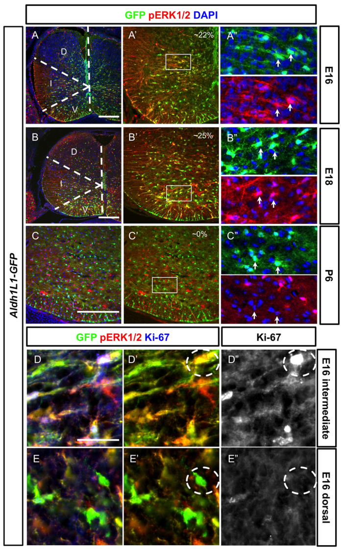
Proliferative Aldh1L1-GFP+ cells in the parenchyma of the spinal cord are often co-labelled with activated ERK1/2. Spinal cord sections from different developmental stages are shown (developmental stages are indicated in the left boxes). (A-C″) Activated pERK1/2 (red) is found in the Aldh1L1-GFP+ cells (green) at E16 (A-A″) and E18 (B-B″) but not at P6 (C-C″). At E16 (A-A″), pERK1/2 is strongly expressed in the intermediate domain (indicated by the dashed line) but not in the dorsal domain whereas strong pERK1/2+ Aldh1L1-GFP+ cells can be identified in all three domains at E18 (B-B″). The colocalisation percentage of pERK1/2+ Aldh1L1-GFP cells among all GFP+ cells is indicated. At P6 (C-C″), expression of pERK1/2 in Aldh1L1-GFP is not observed. The boxed areas in A′-C′ are shown in A″-C″. Arrows indicate GFP+ cells. D, dorsal domain; I, intermediate domain; V, ventral domain. (D-E″) At E16, Aldh1L1-GFP+ cells in the intermediate domain can often be identified with pERK1/2+ and Ki-67+ (D-D″) whereas those in the dorsal domain have not undergone proliferation and express less pERK1/2 and are negative for Ki-67 (E-E″). Compare the dashed circles in D′ and E′. Scale bars: 200 μm in A-C; 40 μm in D.