
Fig. 5.
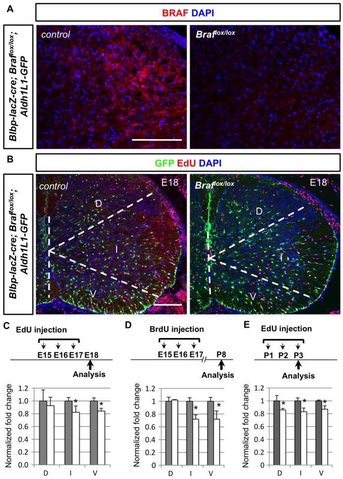
Conditional deletion of Braf results in decrease proliferation of Aldh1L1-GFP+ cells. (A) BRAF expression (red) is largely absent in the Blbp-lacZ-Cre; Braflox/lox spinal cords compared with littermate controls. (B) Decreased number of EdU+ (red) Aldh1L1-GFP+ (green) cells in the Braflox/lox mutant animals compared with littermate controls. (C, D) Animals with EdU or BrdU injection timetable (E15-17) were analysed at E18 or P8, respectively. The percentage of EdU+ Aldh1L1-GFP+ cells among all GFP+ cells is quantified from three different domains, obtained from three independent animals. To obtain normalised fold change, percentage from control animals (grey bars) is set as 1 arbitrarily and a decrease in mutant animals is observed (white bars). Note that the detailed percentage and normalised number is included in supplementary material Table S1. (E) Animals with EdU injection protocols (P1-3) were analysed at P3 and the normalised fold change is shown as describe above. D, dorsal domain; I, intermediate domain; V, ventral domain. Scale bars: 100 μm.