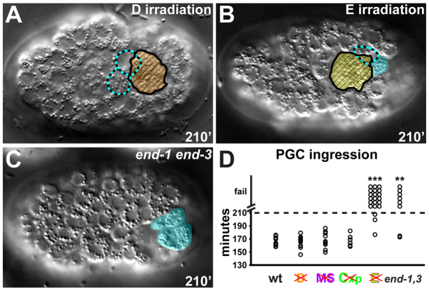
Fig. 2.

Requirement of interacting cells for PGC ingression. (A-C) Embryos (~50 μm in length) are oriented anterior towards the left and are shown from the ventral perspective. PGC ingression following D irradiation (A), E irradiation (B) and in end-1 end-3 mutant embryos (C). PGCs, cyan; E, yellow; D, orange. The position of internalized PGCs is indicated with dashed cyan outlines, while PGCs that failed to ingress are shaded in cyan. The corpse of the irradiated cell remaining on the surface is indicated with hatched fill. (D) Ingression of PGCs in laser-irradiated and mutant embryos (wild-type, n=10; D irradiation, n=12; MS irradiation, n=11; Cxp irradiation, n=6; E irradiation, n=21; end-1 end-3, n=8). Asterisks indicate a significant difference in whether PGCs ingressed relative to unirradiated controls (Fisher's exact test, **P<0.01, ***P<0.001).
