Fig. 6.
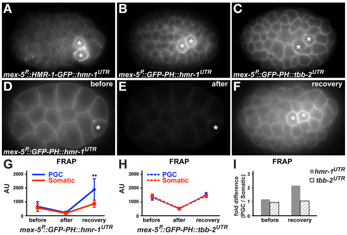
Regulation of HMR-1 enrichment in PGCs. Embryos (~50 μm in length) are oriented anterior towards the left and are shown from the indicated perspective. (A-C) GFP expression from the indicated transgenes. PGCs, asterisks. (D-F) GFP in embryos expressing mex-5P::GFP-PHPLC∂::hmr-1UTR immediately before (D) and after (E) photobleaching, and following recovery (F). Photobleaching was terminated before completion to prevent embryo lethality. (G-I) Quantification of fluorescence recovery after photobleaching (FRAP). AU, arbitrary units; error bars indicate s.d. (mex-5P::GFP-PHPLC∂::hmr-1UTR, n=9; mex-5P::GFP-PHPLC∂::tbb-2UTR, n=7). Asterisks indicate a significant difference in expression levels between PGC and control somatic cell contacts (**P<0.01, two-tailed Student's t-test). (I) Fold difference in expression level between PGC and somatic cell contacts.

