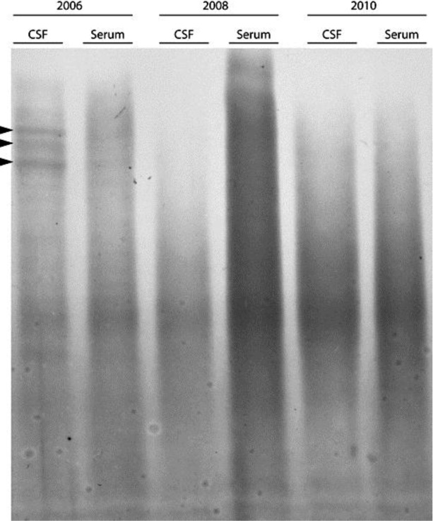
Fig. 1.

Results of isoelectric focusing of IgG in CSF and serum from 2006 (before transplantation) and after transplantation in 2008 and 2010. The anode of the gel is at the bottom of the gel and the cathode is at the top of the gel. Oligoclonal bands are depicted by arrowheads. IgG oligoclonal bands were studied by isoelectric focusing in a commercially available isoelectric focusing gel followed by immunofixation with anti-IgG antibody and protein staining of fixed IgG-anti-IgG complexes in the gel (Hydragel 9 CSF isofocusing, Sebia Electrophoresis, Norcross, USA).
