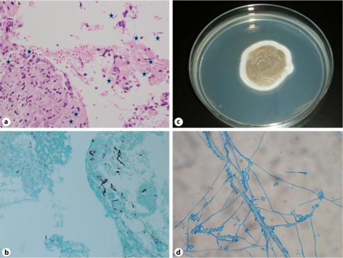
Fig. 2.

The dematiaceous fungi can be identified in the dermis and subcutaneous tissues (a). Grocott staining specimen revealed blackish hyphae throughout the nodules (b). The biopsy material was cultured on Potato Dextrose Agar (PDA) and the gray-blackish villi appeared on the obverse side (c). Slide culture for one week at 25°C. The conidia arising from slightly tapering phialides (d). Hematoxilin and eosin: original magnification ×200 (a), Grocott staining, original magnification ×200 (b).
