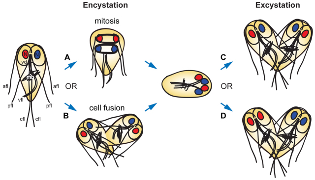
Fig. 1.
Overview of alternative hypotheses for cyst formation and nuclear distribution during excystation. Shown on the left is a diagram of a Giardia trophozoite, with the major components of the microtubule cytoskeleton labeled: the ventral adhesive disc (vd), used for attachment to the intestinal epithelium; four sets of flagella (afl, anterolateral; pfl, posterolateral; vfl, ventral; cfl, caudal); the median body (mb), a microtubule array of unknown function; and the two nuclei (N). Although it has often been assumed that the Giardia cyst is formed after an incomplete mitotic division (A), the alternative route of cell fusion between two trophozoites (B) has never been excluded. In addition, during excystation, two possible routes for the partitioning of nuclei to daughter cells would have dramatically different consequences for Giardia populations. If each daughter cell receives one copy of each parental nucleus (C), any nuclear heterozygosity is maintained throughout the life cycle. By contrast, if the nuclei were distributed such that each daughter cell received two identical nuclei (D), heterozygosity would be eliminated.

