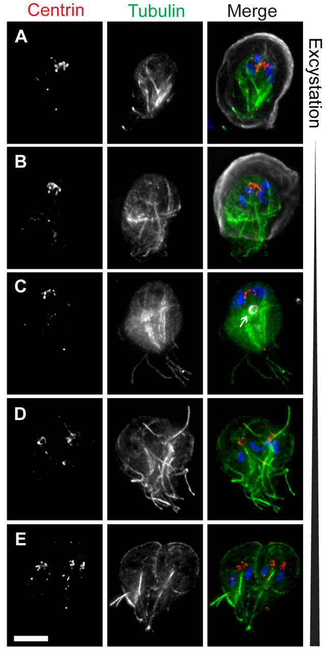
Fig. 3.

Reassembly of the microtubule cytoskeleton during excystation. Excysting cells were stained with DAPI to label DNA (blue) and antibodies to label tubulin (green), centrin (red), and cyst wall protein (CWP) (gray). (A) Cyst in the early stages of excystation (note the rupture in the cyst wall at the bottom of the image). (B) An excyzoite emerging from a cyst. (C) Completely excysted cell. A residual encystation-specific vesicle is indicated (arrow). (D,E) Later-stage excyzoites in which the typical heart-shaped cytokinetic morphology is restored, including two complete discs and 16 flagella. Scale bar: 5 μm.
