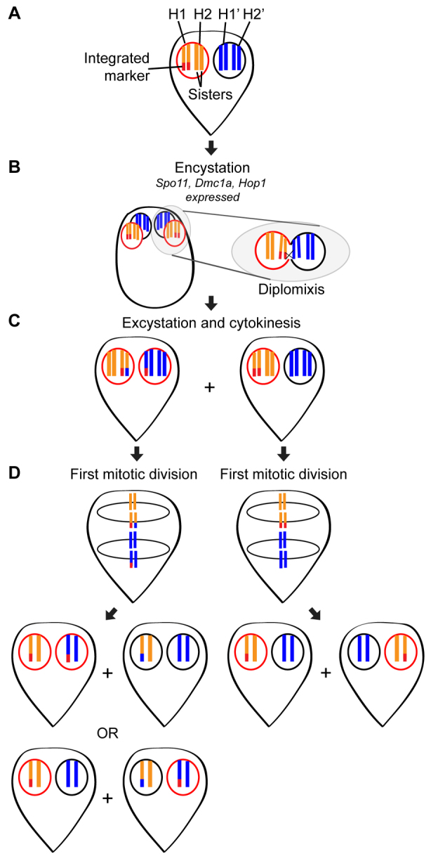
Fig. 6.

Hypothetical model of homologous recombination during diplomixis. (A) Shown is a simplified cell containing an integrated marker (red) in one of its four homologous chromosomes (H1 and H2, shown in orange, in the left nucleus, and H1′ and H2′, shown in blue, in the right nucleus). Throughout the figure, the presence of the integrated marker is indicated by a red outline around the nucleus in which it is found. Because trophozoites typically rest in G2 of the cell cycle, this trophozoite is shown with its chromosomes already duplicated (8C genome content; 4C in each nucleus). (B) During encystation, the meiotic gene homologs Spo11, Dmc1a and Hop1 are induced. Both nuclei divide and then go through an S phase, producing a 16C cyst. At some point, the nuclei fuse and exchange genetic material, presumably through homologous recombination. The chromosomes remain tethered to the nuclear envelope by their telomeres, which might prevent the development of aneuploidy following diplomixis. (C) When the cell excysts and completes cytokinesis, two trophozoites are formed, one from each nuclear pair found in the cyst. (D) If diplomixis has occurred (left trophozoite in C), the first mitotic division produces two possible combinations of daughter trophozoites, depending on the orientation of the chromosomes on the spindle. One of these combinations would produce one cell with markers in both nuclei and one cell with no marker, whereas the other would produce the parental arrangement (marker in one nucleus). If diplomixis has not occurred (right trophozoite in C), only the parental arrangement (marker in one nucleus) is produced.
