
Fig. 3.
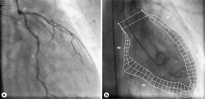
Control cardiac catheterization 2 days later. a Caudal RAO projection of the left coronary artery: normalized left main (bold arrow) and non-significant circumflex artery stenosis (normal arrow), diffusely non-significantly diseased left anterior descending artery (small arrows). b Ventriculography (RAO 30°). Quantitative evaluation revealed a normal left ventricular function (LVEF 60%) and no wall motion abnormalities, with near normal LVEDP of 16 mm Hg.