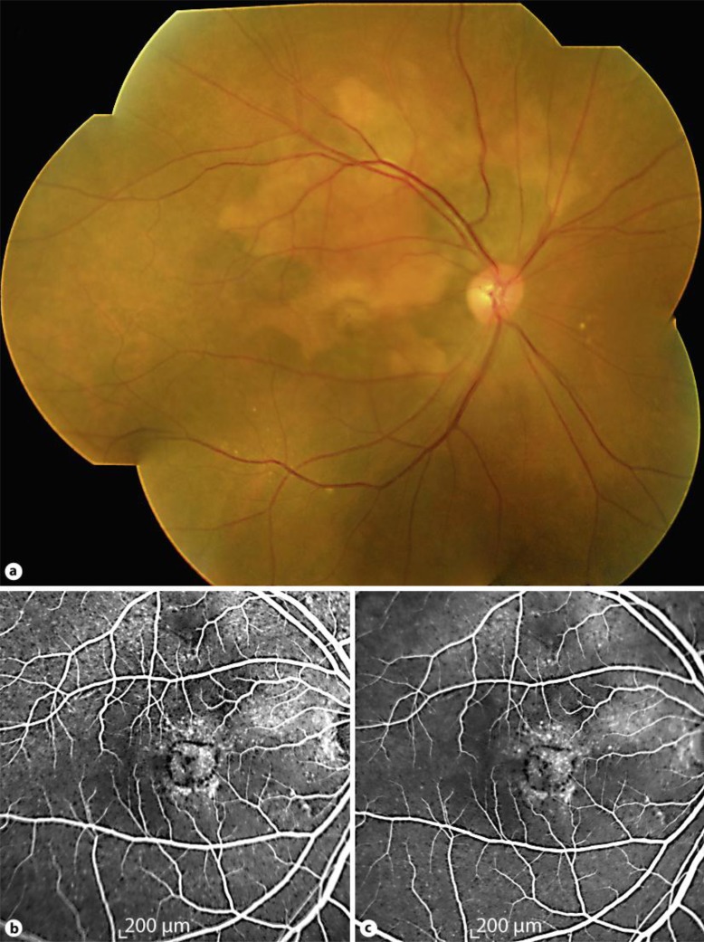
Fig. 2.
a Mosaic fundus photograph of the right eye at 3 months following intravitreal ranibizumab treatment showing complete resolution of macular hemorrhage with mild fibrosis at the inferior border of the choroidal osteoma. b Early- and c late-phase fluorescein angiography showing fibrosis of the choroidal neovascularization with absence of leakage.

