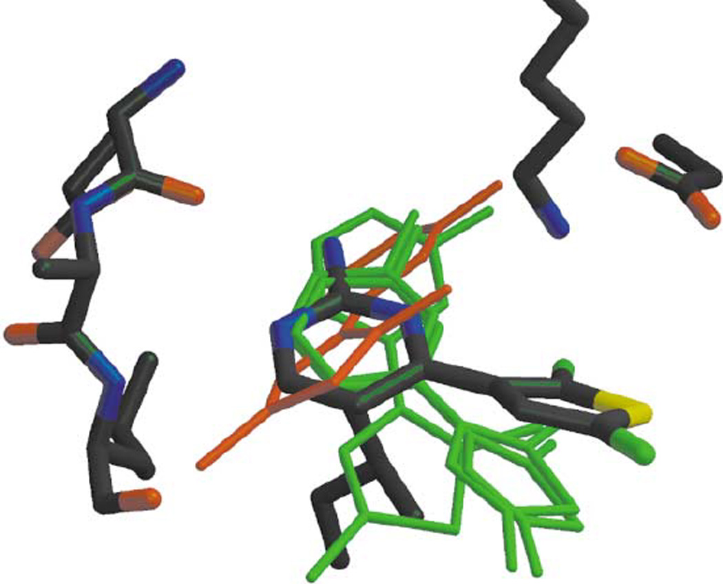
Figure 10.

Overlay of the top four positions identified by the program LIDAEUS, in Green, compared with the experimentally determined X-ray structure of CYC1. The ligand in red is the fourth-best fit. It has an unacceptable energyscore (and large RMSD fit on the experimental structure). (from Ref. [32])
