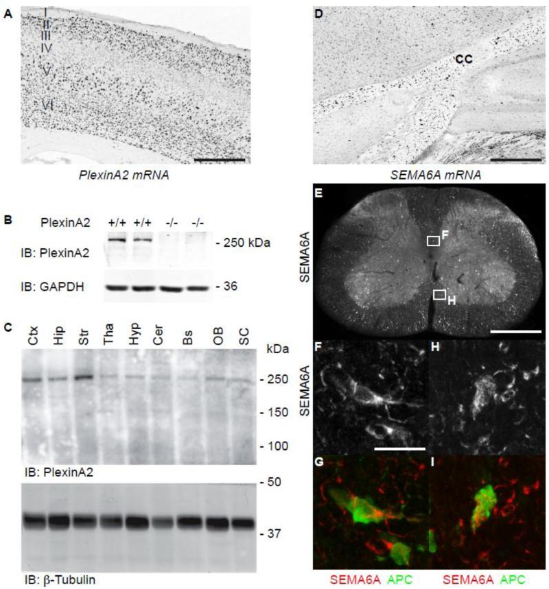
Figure 1. Expression of PlexinA2 and Sema6A in Adult CNS.
Photomicrograph of in situ hybridization demonstrates PlexinA2 mRNA expression is enriched in layer V primary motor and somatosensory cortical neurons in adult wild type mice (A). PlexinA2 expression can also be detected at the protein level in adult wild type but not PlexinA2-/- mice via immunoblot (B). PlexinA2 protein expression is enriched in cortex and striatum, but can be detected throughout the brain and spinal cord (C). Sema6A mRNA is detected in subcortical white matter regions in adult wild type mice, including the corpus callosum (CC) (D). Photomicrographs of transverse cervical spinal sections from adult wild type mice show Sema6A expression in dorsal, lateral and ventral white matter (E). Higher power photomicrographs of dorsal and ventral white matter (insets, F and H), show Sema6A expression in cells morphologically consistent with oligodendrocytes (F, H) identified by co-localization with the mature oligodendroctye marker, APC (G, I). (C, Ctx = cortex, Hip = hippocampus, Str = Striatum, Tha = Thalamus, Hyp = Hypothalamus, Cer = Cerebellum, Bs = brainstem, OB = Olfactory Bulb, SC = spinal cord). Scale bar A, D and E = 500 μm, F = 10 μm.

