
Fig. 2. Ecto/Tif1–γ in early mouse development.
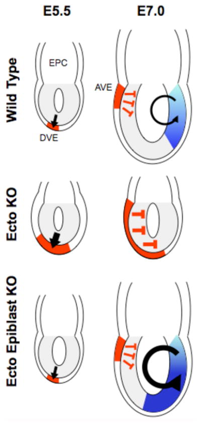
Schematic of mouse embryos with the epiblast (progenitor of the embryo proper) in gray adjacent to extraembryonic cells of the ectoplacental cone (EPC). The thin layer surrounding the epiblast and the EPC is the primitive endoderm. Top row is wild type. At embryonic day 5.5 (E5.5 - one day past uterine implantation) Nodal signaling (black arrow) induces differentiation of the Distal Visceral Endoderm (DVE - in red). At E7.0, one-half day past the initiation of gastrulation, Nodal (black arrow) induces differentiation within the epiblast of mesodermal tissues (shades of blue). Rotation of the DVE toward the anterior forms the Anterior Visceral Endoderm (AVE – in red) that secretes the Nodal antagonists Lefty1 and Cerberus-like (red T-bars). These proteins limit the activity of Nodal within the epiblast. Middle row is Ecto −/−. At stage E5.5. the lack of Ecto/Tif1–γ causes an expansion of the DVE and subsequently the AVE (larger red area). The latter leads to the absence of mesoderm due to production of excess Lefty1 and Cerberus-like (larger red T-bars). Bottom row is Ecto−/− only in epiblast cells. At stage E5.5 these embryos are indistinguishable from wild type. At stage E7.0, AVE expression of Lefty1 and Cerberus-like is wild type but enhanced Nodal signaling (larger black arrow) within the epiblast leads to expansion of mesoderm (larger blue area).