Fig. 5.
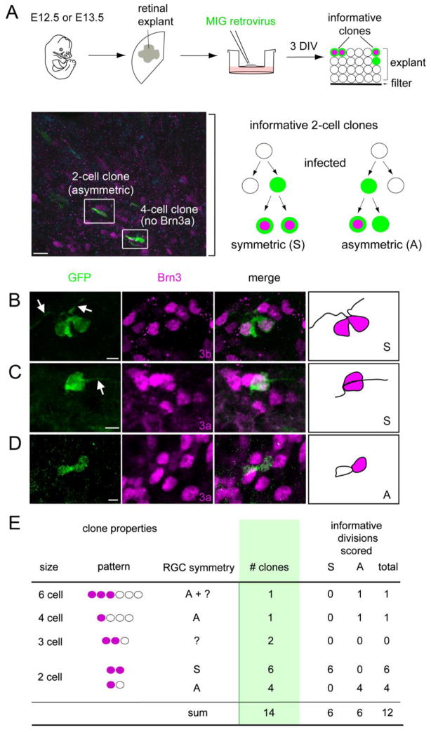
Paired ganglion cells can be generated from retinal progenitors by symmetric terminal division. (A) Retinas were explanted from E12.5 or E13.5 embryos, infected at clonal density with MSCV-IRES-GFP (MIG) retrovirus (green), and cultured for 3 days in vitro (DIV). The micrograph shows a cross-section from a representative explant (bracket) coimmunostained for Brn3a (magenta) and GFP (green). The schema shows informative two-cell GFP+ clones: a symmetric [S] clone containing two Brn3+ (3a or 3b) RGCs, and an asymmetric [A] clone with one Brn3+ RGC. (B–D) Confocal Z-stack projections and drawings from representative symmetric (B, C) or asymmetric (D) clones containing RGCs. Most Brn3+ cells have long processes (arrows), confirming that they are differentiated RGCs. (E) Summary of observed clones containing at least one Brn3+ cell. In these experiments, RGCs were identified using Brn3a (12 clones) or Brn3b (2 clones) antisera interchangeably. Among 12 informative divisions, an equal number with symmetric and asymmetric fates was observed. Scale bars, 10 μm in A; 5 μm in B–D.

