
Figure 6.
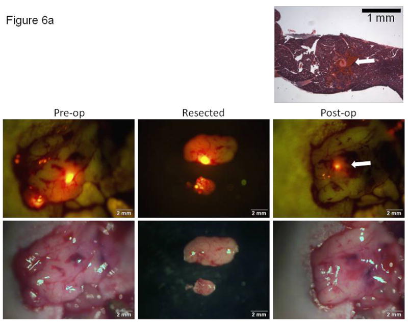
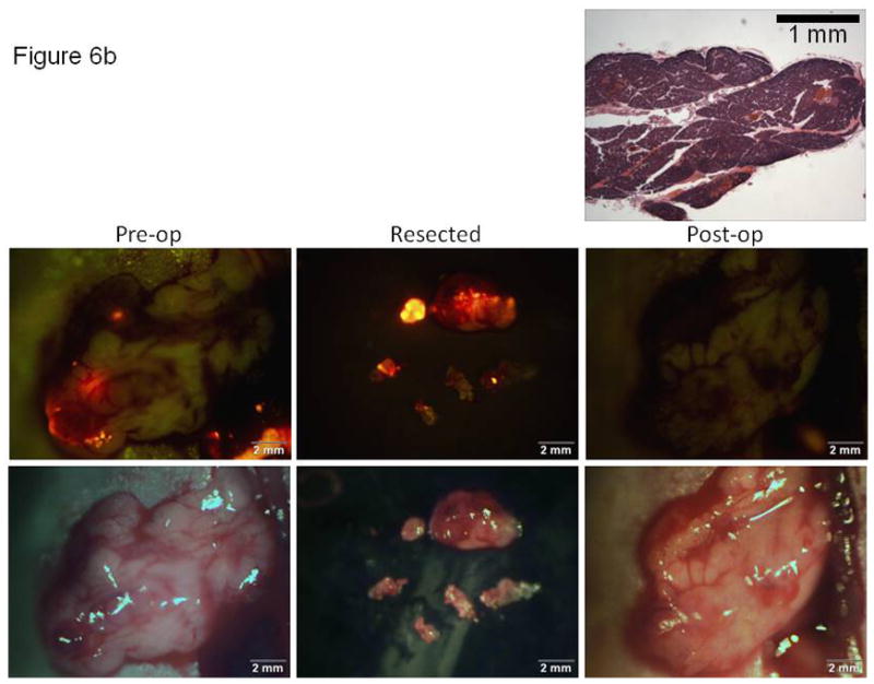
Correlation of imaging with histology for documentation of tumor resection using either bright light or fluorescence-guided surgical techniques. Brightfield (lower) and fluorescence (upper) images obtained at the time of surgery for either (A) a bright light or (B) fluorescence-guided surgical approach. Histologic cross-sections (top image in each 6A and 6B, H&E) taken through the surgical margins of the unresected tissues. The arrows in Figure 6A show the correlation of the small focus of unresected (fluorescent) tumor cells retained in the post-operative tumor bed and the histologic section through this focus in a mouse resected for tumor under BLS.