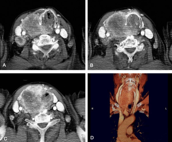
Figure 10.
Contrast enhanced CT scan, axial images and coronal reconstructed image. Axial images sequences show the complete closure of the tracheal lumen. A thyroid mass with substernal extension, and with right jugular vein and carotid artery compression and displacement, in addition to diffuse lymphadenopathy are also evident.

