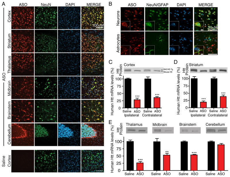
Figure 2. ASOs infused into the cerebral spinal fluid distribute and are active throughout the CNS.
(A and B) An ASO targeting human huntingtin (HuASO) was infused continuously for two weeks into the right lateral ventricle of 2-month-old non-transgenic mice. Immunohistochemical staining of antisense oligonucleotides (red), neurons (NeuN, green), astrocytes (GFAP, green) and nuclei (DAPI, Blue). Antisense oligonucleotides are present in most neuronal nuclei (B, top inset), astrocytes (B, bottom inset) and other non-neuronal cells (examples denoted with *). Scale bar: (A) 100μm, (B) 50μm and inset 5μm. Representative example from two independent experiments, n=4 per treatment.
(C–E) HuASO was infused continuously for two weeks into the right lateral ventricle of 2-month old BACHD mice. Immunoblot of human (upper band) and mouse (lower band) huntingtin, and quantification of human huntingtin mRNA levels (mean % ± SEM relative to saline controls) 8 weeks post-treatment termination in BACHD (C) cortex ipsilateral and contralateral to the injection site, (D) ipsilateral and contralateral striatum (E) thalamus, midbrain, brainstem and cerebellum. Asterisks (*p<0.05, **p<0.01, and ***p<0.001) mark statistically significant changes relative to saline infused animals (n=5 per treatment, two-tailed unpaired t-tests). See also Figure S2.

