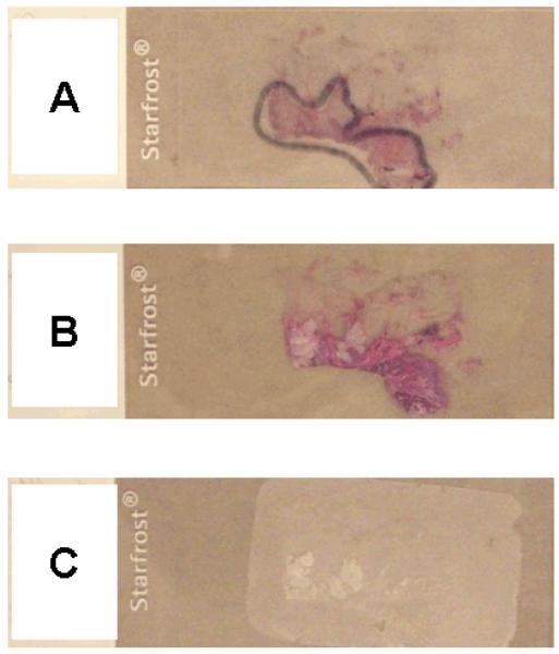
Figure 1.
Examples of HE-stained and unstained tissue sections.
A. A HE-stained tissue section slide to guide tumor tissue dissection. A pathologist marked a tumor area under a microscope.
B. A HE-stained tissue section without coverslip for subsequent DNA extraction. It is easy to identify the tumor area.
C. An unstained tissue section for DNA extraction. It is not as easy to identify the tumor area as in the HE-stained tissue section (B).
HE, hematoxylin and eosin.

