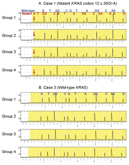
Figure 5.
Tissue staining and subsequent PCR-Pyrosequencing for KRAS
A. Case 1 with mutant KRAS codon 12 (c.35G>A) admixed with wild-type sequence. The assay was repeated 3 times on 120 DNA aliquot specimens (3 cases × 4 groups × 10 sections), and representative results are shown. There was no appreciable difference between DNA specimens from the four groups.
B. Case 3 with wild-type KRAS. The assay was repeated 3 times on 120 DNA aliquot specimens (3 cases × 4 groups × 10 sections), and representative results are shown. There was no appreciable difference between DNA specimens from the four groups.

