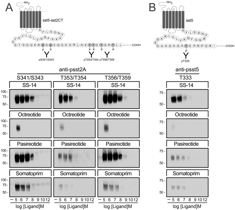
Figure 3. Agonist-selective phosphorylation of the human sst5 and sst5-sst2CT chimera.
(A,Top panel) Schematic representation of the sst5-sst2CT receptor indicating the phosphate acceptor sites S341/343, T353/354 and T356/359 within the carboxyl-terminal tail. (A, Bottom) HEK293 cells stably expressing the sst5-sst2ACT receptor were either not exposed or exposed for 5 min to SS-14, octreotide, pasireotide or somatoprim in concentrations ranging from 10−12 to 10−5 M. The levels of phosphorylated sst5-sst2ACT receptors were then determined using the phosphosite-specific antibodies anti-pS341/pS343 {3157}, anti-pT353/pT354 {0521} and anti-pT356/pT359 {0522}. (B,Top panel) Schematic representation of the human sst5 receptor indicating the phosphate acceptor site T333 within its carboxyl-terminal tail. (B, Bottom) HEK293 cells stably expressing the human sst5 receptors were either not exposed or exposed for 5 min to SS-14, octreotide, pasireotide or somatoprim in concentrations ranging from 10−12 to 10−5 M. The levels of phosphorylated sst5 receptors were then determined using the phosphosite-specific antibodies anti-pT333 {3567}. Western blots shown are representative of three to five independent experiments for each condition. The positions of the molecular mass markers are indicated on the left (in kDa).

