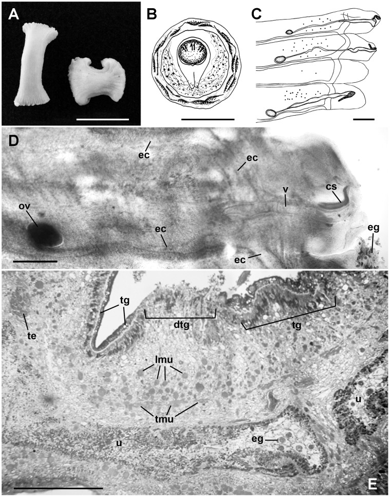
Figure 1. Morphological features of Bertiella sp. expelled by a 4-year-old girl of the Amazon region.
(A) Macroscopic view of proglottids (Bar = 1 cm). (B) Egg illustration showing thick shell, albuminous layer, piriform apparatus, and oncosphere (Bar = 2.5 µm). (C) Mature proglottids illustration showing genital pores in an irregular, alternating arrangement, unlobed ovaries, vagina, cirrus sac, and excretory canals (Bar = 1000 µm). (D) Gravid proglottids under compression showing the ovary (ov), vagina (v), cirrus sac (cs), and excretory channels (ec). Numerous eggs (eg) were observed outside of proglottids (Bar = 500 µm). (E) Proglottids histological section, showing few eggs (eg) in the uterus (u). The tegument presented intact areas (tg) and discontinuous portions (dtg). Transversal (tmu) and longitudinal (lmu) muscles and testes (te) were also observed (Bar = 200 µm).

