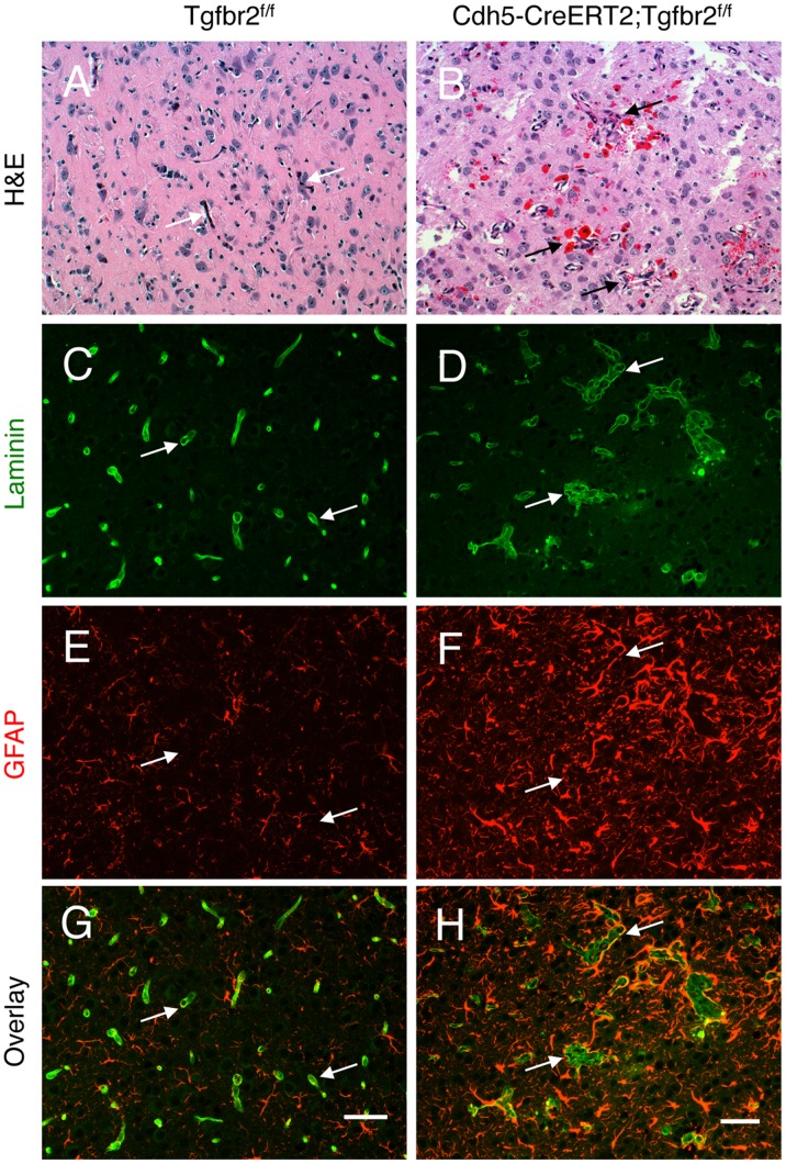
Figure 1. Loss of Tgfbr2 in neonatal endothelial cells leads to cerebral vascular pathologies and intracerebral haemorrhage.
Coronal sections through cerebral cortices of P14 control (A) and Tgfbr2–iKOe mice (B) were stained with H&E. Note the abnormal blood vessel morphologies and microhaemorrhage in Tgfbr2 mutant brains. (C-H); Coronal sections through cerebral cortices of P14 control (C, E, G) and Tgfbr2 conditional mutant mice (D, F, H) were double immunofluorescently labelled with anti-laminin (C, D) and anti-GFAP (E, F) to visualize vascular basement membranes and astrocytes, respectively. Note the cerebral blood vessels with glomeruloid-like tufts (D) as well as robust perivascular astrogliosis (F) in Tgfbr2-iKOe mutant sections. A total of 5 mutant and 4 control mice were examined for brain pathologies. Scale bars: 50 µm.

