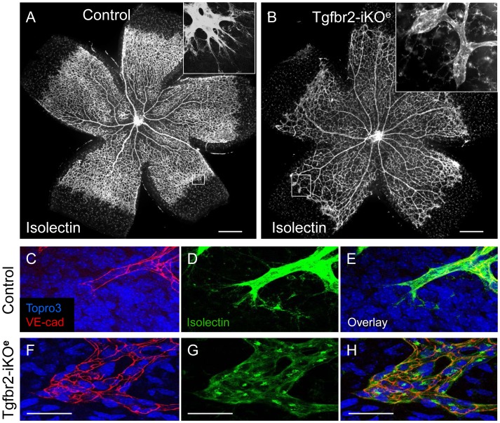
Figure 2. Tgfbr2-iKO mutants show vascular abnormalities in the postnatal retina.
Isolectin stained retinal preparations at postnatal day (P)7 show normal vascular architecture in controls (A), but reduced branching in Tgfbr2-iKOe mutants (B). Frequently, round clusters of endothelial cells (boxed area and inset, B) are seen at the leading edge of the vascular plexus in mutants, at the positions where tip cells normally occur in controls (boxed area and inset, A). High power views of whole mount P9 retinal preparations stained with VE-cadherin and isolectin show the endothelial footprint of a normal tip cell (C,D,E) compared with that of the endothelial cell clusters in the Tgfbr2-iKOe mutant (F,G,H). Scale bars: 500 µm A,B; 50 µm, C–H.

