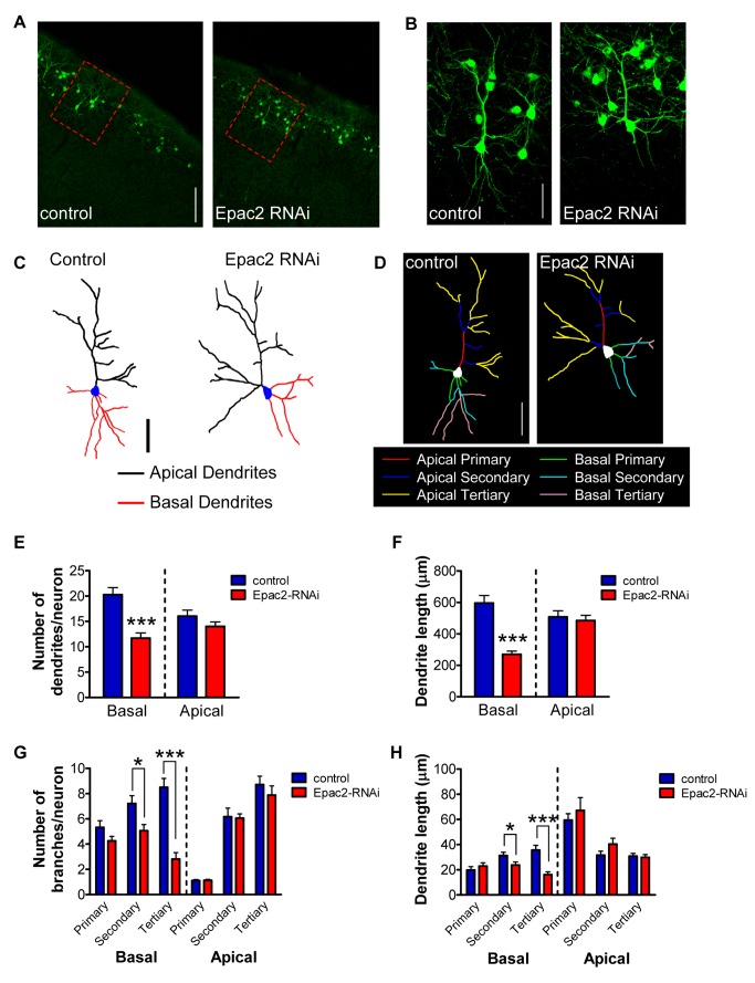
Figure 1. Epac2 regulates basal dendrite complexity in vivo.
(A) Low magnification images of in utero electroporated layer 2/3 cortical neurons, expressing either control or Epac2-RNAi. Animals were electroporated at E16.5; coronal slices (50 µm) were made at P28. Red dashed rectangles indicate images represented in (B–D). (B) High magnification images of layer 2/3 cortical neurons outlined in (A). (C–D) Binary images (C) and “skeleton outline” of basal and apical arbors (D), separated into primary, secondary, and tertiary branches, of neurons shown in (B). (E) Quantification of dendrite numbers separated into total apical and basal dendrite branches. Epac2 knockdown in vivo induced a loss of basal dendrites. (F) Epac2 knockdown in vivo induced a selective reduction in basal dendrite length. (G) Quantification of dendrite branch numbers separated into basal/apical, primary/secondary/tertiary order branches. Epac2 knockdown in vivo induced a loss of basal dendrites, specifically driven by a loss of secondary and tertiary basal dendrites. (H) Epac2 knockdown in vivo selectively reduced secondary and tertiary basal dendrite lengths. *p<0.05, ***p<0.001; scale bars, 100 µm (A); 50 µm (B–D).

