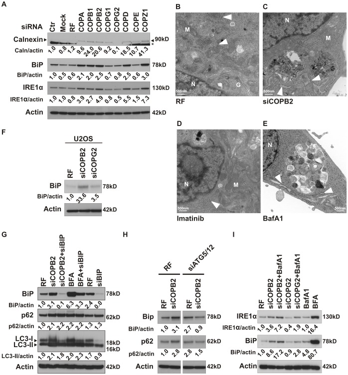
Figure 6. Loss of coatomer subunits promotes ER stress and activates UPR.
(A) Independent COPI subunits were depleted in MDA-MB-231 cells and levels of calnexin, BiP, and Ire1α were measured by westernblot after 72 h. *, a non-specific band. (B–E) Transmission electron microscopy analysis of MDA-MB-231 cells after 48 h treatment with non-targeting siRNA (RF) (B), siCOPB2 (C) or overnight treatment with imatinib (10 µM) (D) or BafA1 (50 nM) (E). Electron micrograph showing the dilated ER (white arrow heads in C and E). M: mitochondria, N: nucleus and G: Golgi apparatus. (F) COPB2 or COPG2 depleted U2OS cells were analyzed for BiP levels. (G) LC3, p62 and BiP levels were analyzed by immunoblot after depletion of COPB2 and BFA treatment with or without BiP siRNA. (H) MDA-MB-231 cells were incubated with control siRNA or COPB2 siRNA, either alone or in combination with siRNA targeting the Atg5/Atg12 complex. Images from different parts of one gel were grouped. (I) MDA-MB-231 cancer cells were depleted of COPB2 or COPG2, either alone or with BafA1, and Ire1α and BiP levels were analyzed by immunoblot. BFA was used as a positive control for ER stress.

