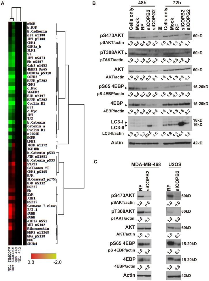
Figure 7. RPPA analysis identifies signaling pathways altered during abortive autophagy.
(A) RPPA analysis was performed on lysates of MDA-MB-231 cells depleted from COPB2 for 48 h or 72 h. The heatmap represents values of triplicates normalized to a pool of controls (untransfected, mock transfected and RF transfected cells). Significant changes in proteins related to depletion of COPG2 were excluded. (B,C) Phosphorylated and total levels of the indicated proteins were validated in MDA-MB-231, MDA-MB-468 and U2OS cells. Actin was used as a loading control. M: marker.

