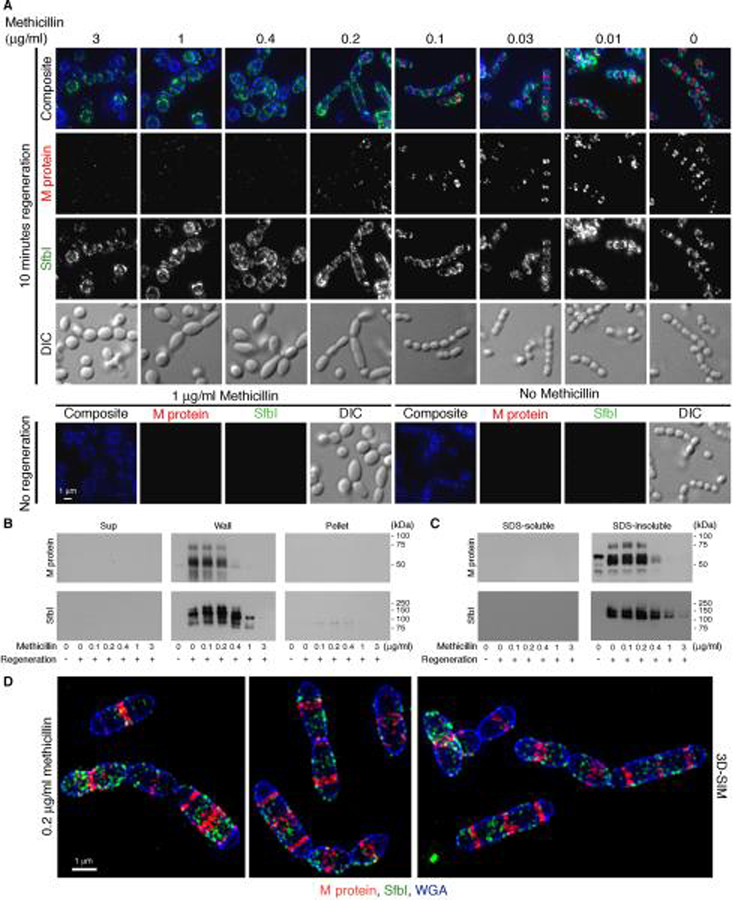
Figure 5. Methicillin-induced unbalanced peptidoglycan synthesis results in a substantial reduction in the cellular amount of M protein compared to SfbI.
An overnight S. pyogenes D471 culture was diluted 1:100 into TH+Y containing trypsin and pronase, and grown at 37°C to OD600 0.5. The cells were then diluted 1:4 into tubes containing TH+Y, trypsin, pronase, and ascending concentrations of methicillin. Following one hour, the cells were washed, and resuspended for 10 minutes in TH+Y containing a similar concentration of methicillin but lacking proteases, and then fixed. (A) M protein (red) and SfbI (green) were labeled using specific antibodies, and the cell wall was stained with WGA marina blue (blue). Deconvolution immunofluorescence images are presented as maximum intensity projections. (B) Similar cultures were fractionated into supernatant, wall, and spheroplast pellet, and processed by Western blot. (C) Additional cultures were harvested, boiled in 2% SDS, and separated into supernatant (“SDS Soluble” fraction), and cell pellet (“SDS insoluble” fraction), which was subsequently treated with the phage lysin PlyC to release wall-anchored proteins, prior to processing by Western blot. (D) Cells treated with 0.2 μg/ml methicillin, were visualized by 3D-SIM and are presented as maximum intensity projections.

