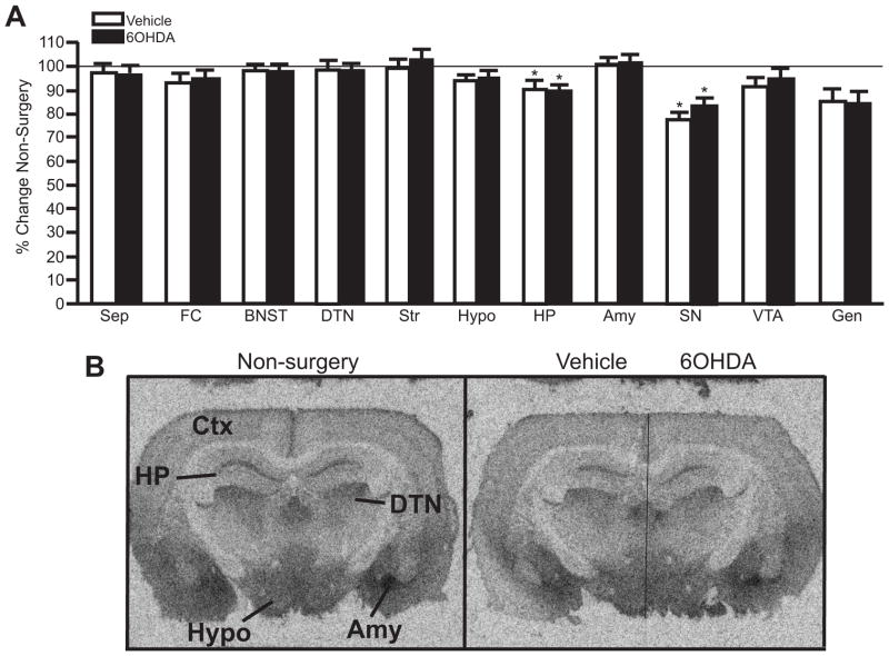
Figure 7.
Unilateral LC 6OHDA does not alter α2-AR binding sites in forebrain regions compared to vehicle-treated side. A) Quantitation of α2-AR binding sites in forebrain regions of non-surgery animals and vehicle/6OHDA-treated animals as a percentage of non-surgery animals. B) Photomicrographic images of α2-AR binding at the level of the HP in non-surgery animal (left panel) and vehicle/6OHDA-treated animal (right panel). Horizontal line on vehicle/6OHDA image denotes midline. * Indicates significant difference from non-surgery animals. Sep, septum; CTX, cortex; FC, frontal cortex; BNST, bed nucleus of the stria terminalis; DTN, dorsal thalamic nucleus; Str, striatum; Hypo, hypothalamus; HP, hippocampus; Amy, amygdala, SN, substantia nigra; VTA, ventral tegmental nucleus; Gen, geniculate.

