Figure 1.

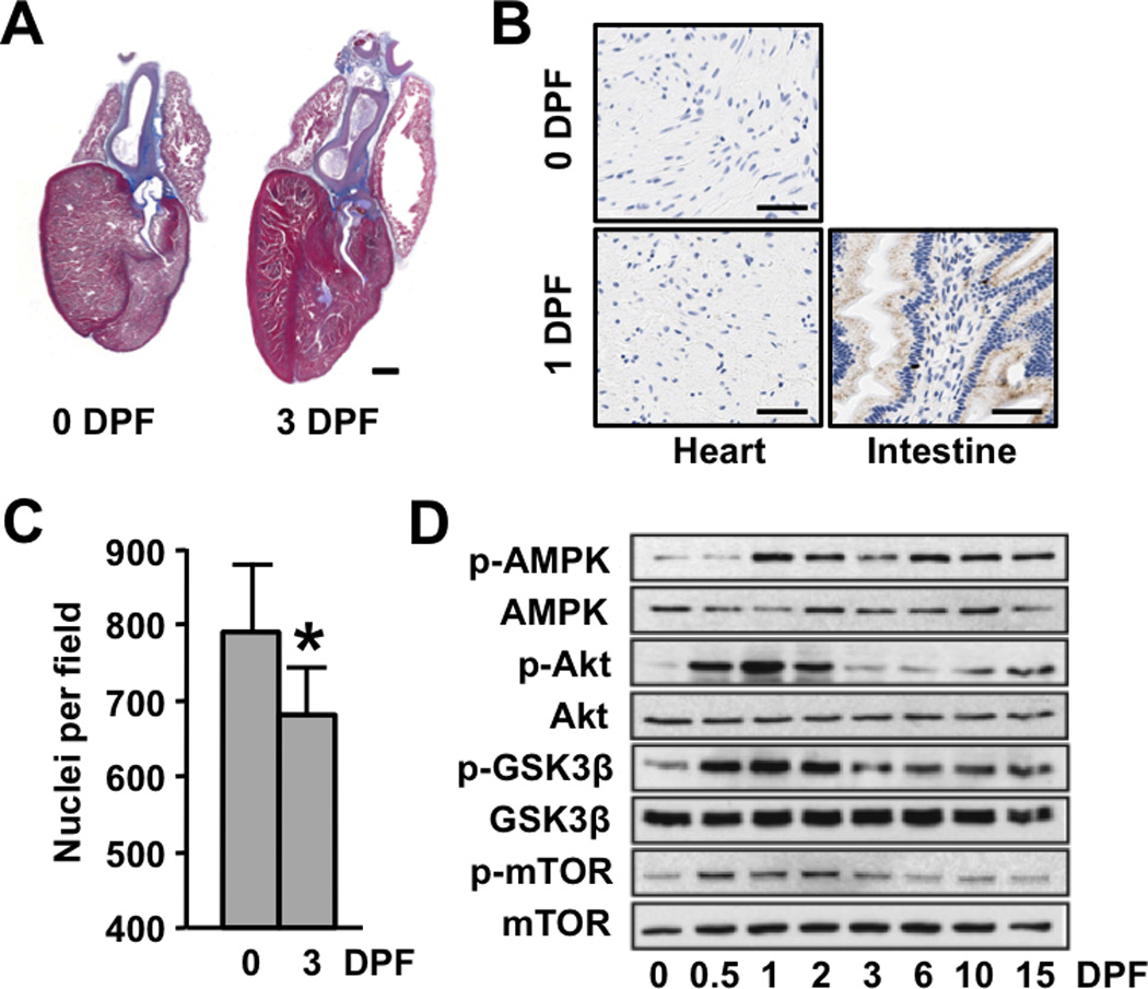
Postprandial cardiac growth in the python is characterized by cellular hypertrophy and activation of protein synthesis pathways. (A) Masson trichrome–stained python hearts depicting pronounced postprandial cardiac hypertrophy. Scale bar = 2 mm (B) BrdU-staining of 0 and 3 DPF python hearts shows no evidence of postprandial cellular proliferation (python small intestine is included as a positive control [brown nuclear staining]) Scale bar = 50 µm (C) The number of nuclei per field is reduced post-feeding. (D) Immunoblot analysis reveals increased phosphorylation of AMPK, Akt, GSK3β, and mTor in the postprandial python heart. Error bars represent ±SE; n=4 per condition; *p<0.05 versus 0 DPF.
