
Figure 2.
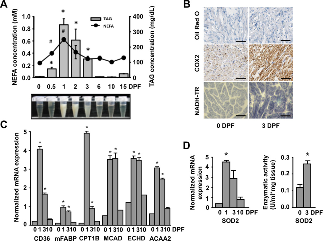
The postprandial python heart has increased expression of fatty acid transport, handling, and oxidation genes along with enhanced free radical scavenging capacity. (A) Plasma fatty acid (FA) and triacylglyceride (TAG) content is significantly increased post-feeding (B) Oil Red-O staining reveals no cardiac accumulation of neutral lipids at 3 days after feeding. Mitochondrial staining is increased in the post-fed python heart as determined by cytochrome c oxidase II (COX2) immunostaining and NADH-tetrazolium reductase (NADH-TR) histochemistry. Scale bar = 50 µm. (C) There is increased mRNA expression of fatty acid transport protein (CD36), muscle-type fatty acid binding protein (mFABP), carnitine palmitoyltransferase (CPT1B), and the β-oxidation genes medium chain acyl-CoA dehydrogenase (MCAD), peroxisomal enoyl-CoA hydratase (ECHD) and acetyl-CoA acyltransferase 2 (ACAA2) post-feeding. (D) The mRNA expression and activity of mitochondrial superoxide dismutase 2 (SOD2) is increased post-feeding. Error bars represent ±SE; n=4 per condition; * and # p<0.05 versus 0 DPF.