
Figure 3.
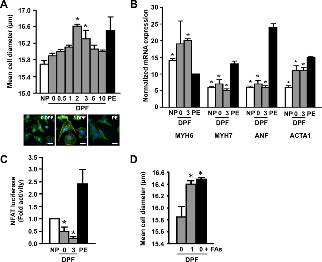
Postprandial python plasma induces cardiomyocyte growth in vitro. (A) Fed python plasma induces cellular hypertrophy in neonatal rat cardiac myocytes (NRVMs). Scale bar = 10 µm. (B) Python plasma does not induce the mRNA expression of known cardiac stress markers in NRVMs. PE, phenylephrine (included as a positive control); ANF, atrial natriuretic factor; MYH6, α-myosin heavy chain; MYH7, β-myosin heavy chain; ACTA1, α-skeletal actin. (C) Pathological NFAT signaling is repressed by python plasma. (D) Supplementing fasted python plasma with C14:0, C16:0, and C16:1n7 (0 DPF + FAs) results in cellular hypertrophy comparable to that seen with 1 DPF plasma. Error bars represent ±SE; n=3 per condition; *p<0.05 versus 0 DPF (A and D); *p<0.05 versus PE (B); *p<0.05 versus no plasma (NP, C).